SEM Viewing Modes in Materials Analysis

Changing how we look at things

SEM Viewing Modes in Materials Analysis

In the investigation of the failure of materials and in materials analysis, we use several analytical methodologies to elucidate facts. One method which we have previously, discussed, is Scanning Electron Microscopy (SEM). SEM is an incredibly powerful tool for any forensic materials engineer, which offers our scientists a method to capture in intimate detail, high resolution photographs at high magnification. SEM can be used in many subtly different ways, and today we will discuss one of those: the viewing modes.

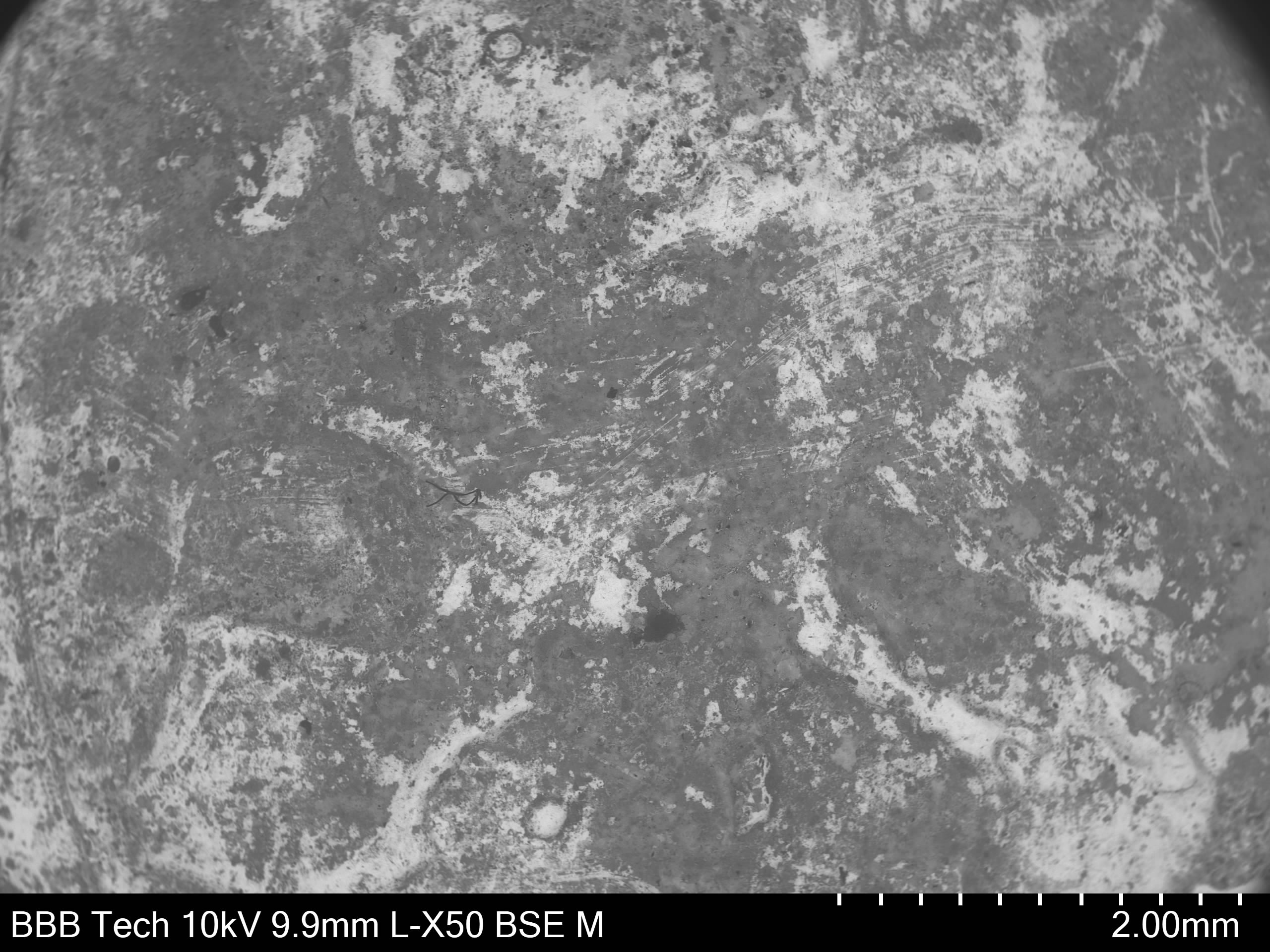
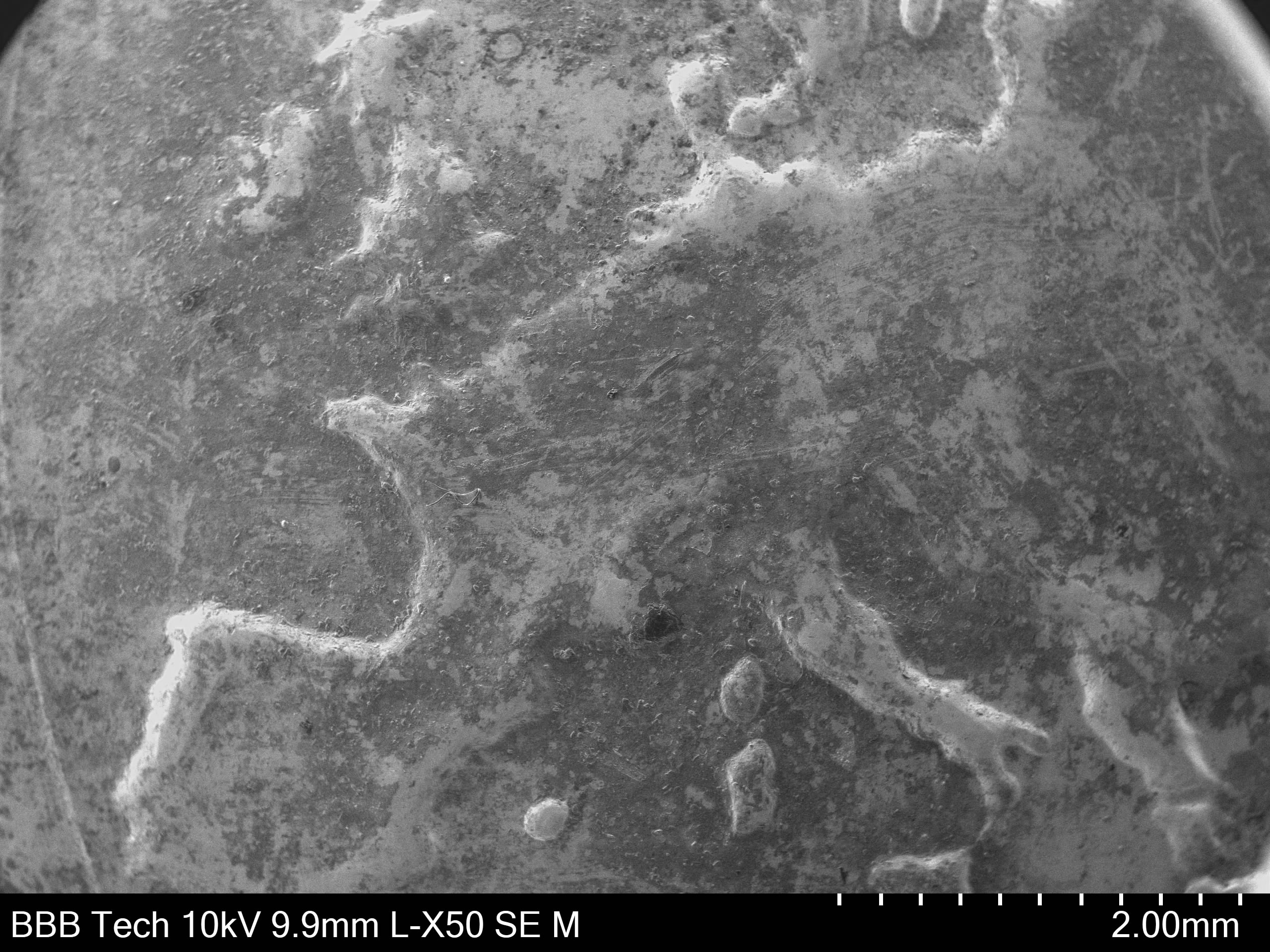
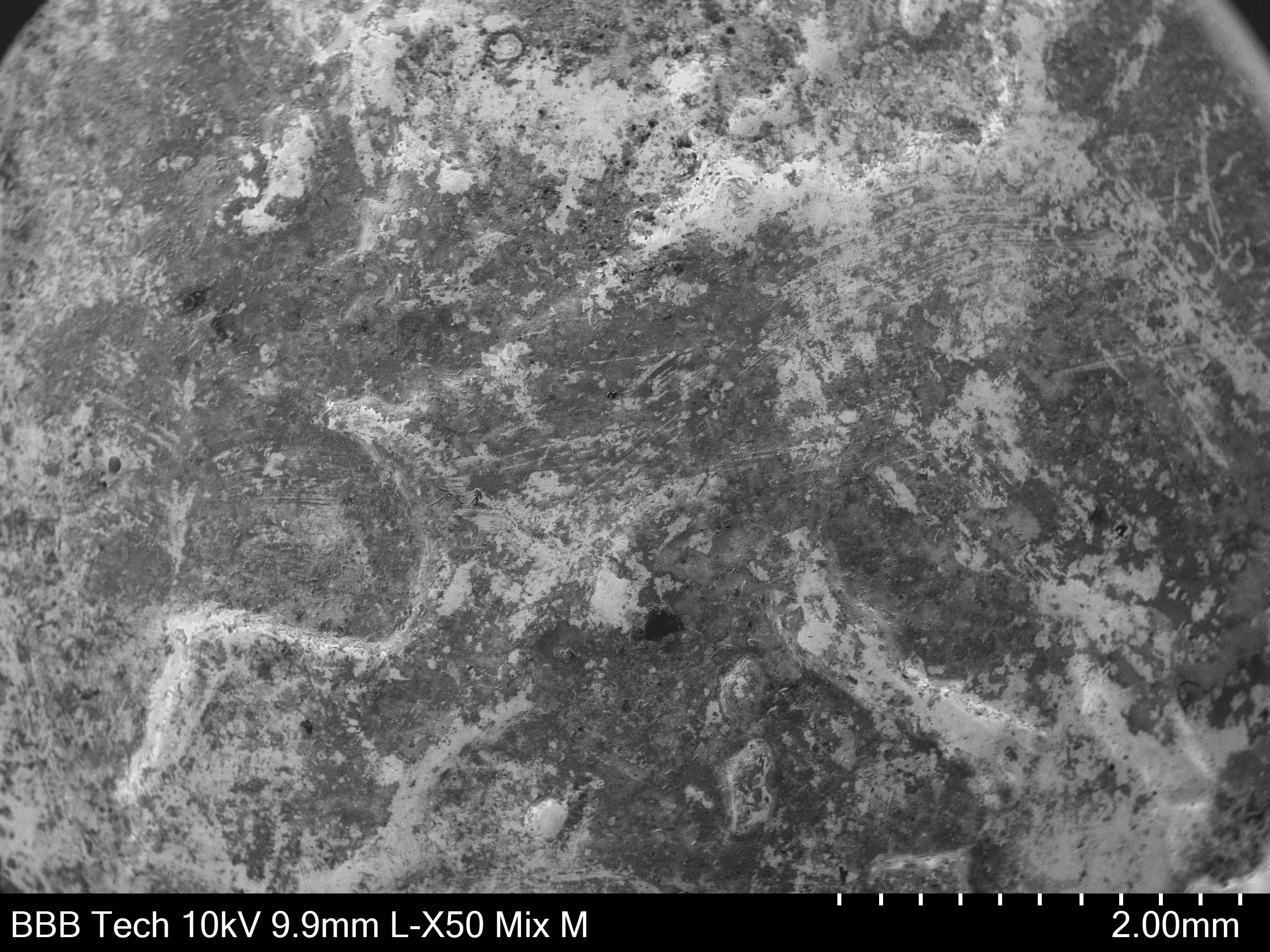
To understand viewing modes, we first need to understand the basics of how SEM works. Essentially, a beam of electrons is generated and shot at the sample, scanning back and forth over the surface. Our Hitachi TM4000Plus SEM has three different viewing modes:

  1. BSE (Back Scattered Electrons)
  2. SE (Secondary Electrons)
  3. Mixed

The effects of the different viewing modes can be seen through the photographs below, which all depict the same surface. 

Back Scattered Electrons viewing mode

Back Scattered Electrons viewing mode


Some of the generated electrons will actually collide with the atoms of the sample, and are bounced back. These bounced back, or "back scattered", electrons are detected by the SEM. The amount of detected back scattered electrons is used to create a greyscale image. Denser elements are more likely to back scatter an electron, and thus will appear brighter/whiter in the image. As we see in the image above, the surface is quite dirty, with many different elements present on the surface (to find out what elements these are, we would use Energy Dispersive X-ray Spectroscopy). However, we don't see much topographical detail to know what we are actually looking at.

Secondary Electrons viewing mode

Secondary Electrons viewing mode


Some of the generated electrons, instead of colliding directly with the atoms of the sample, will cause electrons to ejected from the sample. These are known as secondary electrons. Surface edges will produce more secondary electrons than a flat surface, giving us much more topographical detail. The SE viewing mode shows us that we're actually looking at the map of Europe on a 10 cent coin! However, we have lost any of the elemental differentiation provided by BSE, so we cannot tell the condition of the surface of the coin. 

Mixed viewing mode

Mixed viewing mode

Mixed viewing mode uses both BSE and SE to compose the image, providing us with both elemental and topographical information, and is the go-to mode for initial examination of a sample. While mixed viewing mode images can be a little bit confusing (in the above image, it is difficult to distinguish the edges of Ireland due to the elemental differences), the information provided allows for quick examination of the condition of a sample. The viewing mode can be refined from there as required. 

This is part of why SEM is such a powerful tool. Using the same instrument, we can examine a sample for topographical abnormalities or elemental contamination simultaneously. Do you have a sample to be examined under SEM? Then contact us today!

Our business is solving problems, so talk to us now.

Contact us